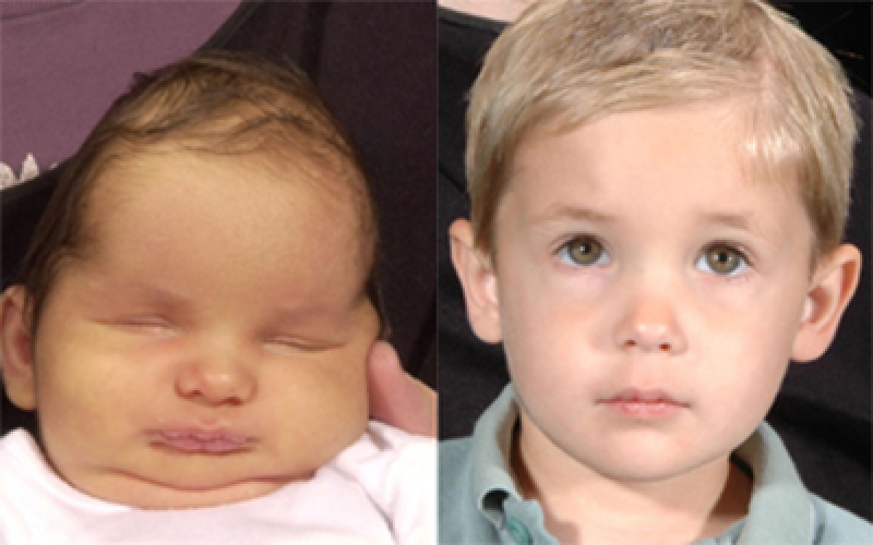
What Is Craniosynostosis?
Craniosynostosis is a condition in which one or more of the fibrous sutures in an infant skull prematurely fuses by turning into bone (ossification), thereby changing the growth pattern of the skull.
Sometimes the resulting growth pattern provides the necessary space for the growing brain, but results in an abnormal head shape and abnormal facial features.
Craniosynostosis results in increased intracranial pressure leading possibly to visual impairment, sleeping impairment, eating difficulties, or an impairment of mental development combined with a significant reduction in IQ.
Moreover, facial deformities associated with craniosynostosis may result in speech and language problems and poor self-esteem.
Craniosynostosis is part of a syndrome in 15 to 40% of the patients, but it usually occurs as an isolated condition.
Causes Of Craniosynostosis:
Craniosynostosis may be caused by a number of factors. They include:
- Biomechanical factors
Due to fetal head constraint - Environmental factors
Maternal smoking
Maternal exposure to amine-containing drugs - Hormonal factors
Hyperthyroid induced craniosynostosis is a hormone mediated premature closure - Genetic factors
Apert syndrome
Pfeiffer syndrome
Crouzon syndrome
Symptoms Of Craniosynostosis:
Signs of craniosynostosis include:
- A misshapen skull, with the shape depending on which of the cranial sutures are affected
- An abnormal feeling or disappearing “soft spot” (fontanel) on the baby’s skull
- Slow or no growth of the head as the baby grows
- Development of a raised, hard ridge along affected sutures
- Increased pressure within the skull (intracranial pressure)
Diagnosis Of Craniosynostosis:
Craniosynostosis can be diagnosed via:
- A physical exam.
Examination of the baby’s head for abnormalities such as suture ridges and facial deformities. - Imaging studies.
A computerized tomography (CT), may show whether any sutures have fused.
X-rays also may be used to measure precise dimensions of the baby’s skull, using a technique called cephalometry. - Genetic testing.
Treatment Of Craniosynostosis:
While mild cases may not need any form of treatment, severe cases may need surgery in order to treat craniosynostosis.
Surgical options can be further divided into:
- Traditional surgery.
This involves reshaping the affected portion of the skull.
Plates and screws are used to hold the bones in place.
In case of recurrence, a second surgery may be required. - Endoscopic surgery.
This involves using a lighted tube (endoscope) inserted through one or two small scalp incisions over the affected suture.
The suture is then opened, to enable the baby’s brain to grow normally.
By : Natural Health News