Bladder Cancer: Three Types and Treatments

Bladder Cancer
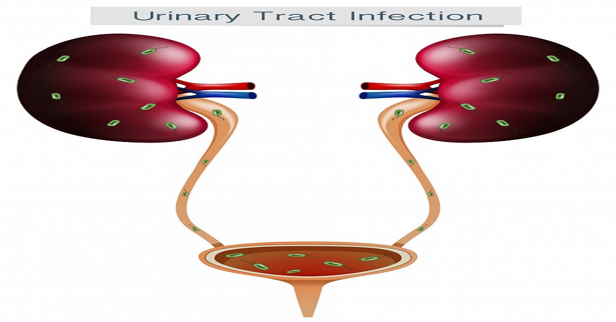
Bladder cancer growth is a typical sort of disease that starts in the cells of the bladder. The bladder is an empty solid organ in your lower midsection that stores waste collected by the kidneys in the form of urine.
Bladder cancer growth regularly starts in the cells, urothelial cells that line within your bladder. Urothelial cells are likewise found in your kidneys and the cylinders (ureters) that interface the kidneys to the bladder. Urothelial malignancy can occur in the kidneys and ureters, as well, yet it’s considerably more typical in the bladder.
Most bladder malignant growths are analyzed at a beginning phase, when the disease is profoundly treatable. Yet, even beginning phase bladder diseases can return after effective treatment. Consequently, individuals with bladder disease ordinarily need follow-up tests for quite a long time after therapy to search for bladder malignant growth that repeats.
Transitional Cell Carcinoma
The tube that associates the kidneys to the bladder is known as the ureter. Most healthy individuals have two kidneys and, in this manner, two ureters.
The highest point of every ureter is found in the kidney in a space known as the renal pelvis. Urine gathers in the renal pelvis and is depleted by the ureter into the bladder.
The renal pelvis and the ureter are fixed with certain kinds of cells called temporary cells. These cells can twist and stretch without falling to pieces. Malignant growth that starts in the temporary cells is the most well-known sort of disease that creates in the renal pelvis and ureter.
At times, temporary cell malignant growth metastasizes, which implies that disease from one organ or some portion of the body spreads to one more organ or part of the body.
Squamous Cell Carcinoma
Squamous cell carcinoma is the second most normal sort of urinary cancer. It represents around 5% of bladder tumors. This disease starts in the flimsy, level squamous cells that might frame in the bladder after constant pain and contamination. Squamous cell carcinoma is frequently found in regions of the world where a parasitic contamination called schistosomiasis is highly prevalent
Adenocarcinoma
Adenocarcinoma is a very rare type of cancer. It generally starts after the glandular cells formation inside the bladder after the persistent irritation and inflammation. Glandular cells are the ones that comprise into mucus secreting cells in the human body.
Spread of bladder cancer
The wall of the bladder has numerous layers. Each layer is composed of various types of cells.
Most bladder tumors start in the deepest covering of the bladder, which is known as the urothelium or transitional epithelium. As the malignant growth develops into or through different layers in the bladder divider, it has a higher stage, turns out to be further developed, and can be more complex to treat.
Over the long haul, the urinary bladder cancer growth may develop outside the bladder and into neighboring constructions. It may spread to local lymph hubs, or to different pieces of the body. At the point when bladder malignant growth spreads, it will in general go to far off lymph nodes, the bones, the lungs, or the liver.
Diagnosis of the Bladder Cancer
Your PCP will need to investigate your urine (urinalysis) to decide whether a contamination could be a reason for your side effects. A minuscule assessment of the urine, called cytology, will search for malignant growth cells.
A cystoscopy is the primary methodology to recognize and analyze bladder disease. In this technique, a lit telescope (cystoscope) is embedded into your bladder from the urethra to see within the bladder and, when done under sedation, take tissue tests (biopsy), which are subsequently analyzed under a magnifying instrument for indications of disease. At the point when this technique is done in the urology specialist’s office, nearby numbing gel is put into the urethra preceding the method to limit the inconvenience.
In the event that the determination of bladder disease is made, the following stage is to eliminate the cancer for clear and detailed arranging and finding.
Transurethral resection (TURBT) is a technique done under broad or spinal sedation in the working room. A telescope is embedded into the bladder and the growth is eliminated by scratching it from the bladder divider (a part of the bladder divider is taken out with it), utilizing an extraordinary cystoscope (called a resectoscope). This technique is demonstrative just as restorative.
This regularly should be possible as a short term system, with patients released from the clinic that very day. After evacuation, the growth is examined by a pathologist, who will decide the sort of cancer, the cancer grade and the depth of attack. The motivation behind the method is to eliminate the cancer and acquire significant organizing data, for example, the growth grade and profundity of invasion.
For certain patients with invasive malignant growth, a CT sweep of the mid-region and pelvis may be the following stage to decide whether there is any spread of the infection outside of the bladder.
Magnetic resonance imaging, which utilizes a magnet, radio waves, and a PC to take definite pictures, should likewise be possible, and is useful in arranging bladder cancer treatment.
A chest X-ray may likewise be performed to recognize if any malignant growth has spread to the lungs. On occasion, a bone scan might be performed to search for metastasis (spread) of the malignant growth deep down. The majority of these tests are utilized specifically, i.e., just in specific patients with related symptoms.


