What is Zygomycosis?
Zygomycosis is a rare infection caused by a class of fungi called Zygomycetes. These are primitive, fast growing, terrestrial, largely saprophytic fungi with a cosmopolitan distribution and live on decaying organic matter.
There are 2 orders of Zygomycetes:
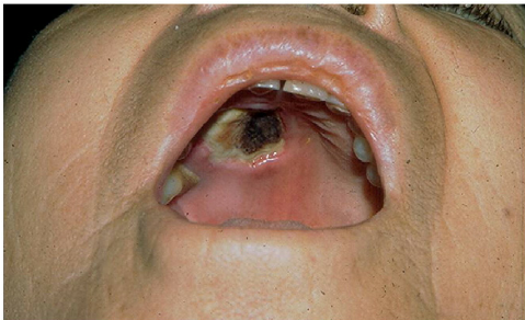
Mucorales are rapidly growing fungi including two families, the Mucoraceae and Cunninghamellaceae. Mucorales usually causes infection in individuals with compromised immune systems due to drugs such as systemic steroids and diseases such as lymphoma and poorly controlled diabetes. The fungi invade blood vessels and cause mucormycosis, an acute, rapidly spreading and fulminant systemic mycosis Rhinocerebral (nose and brain), lung, gastrointestinal and abdomen pelvic, cutaneous and widespread forms have been reported. The mortality rate is very high. Cutaneous lesions from Mucorales are due to traumatic implantation or secondary to spread via the bloodstream to the skin. Mucorales infection may result in plaques, pustules and abscesses or necrotic, ulcerated lesions. The most common species to cause zygomycosis is Rhizopus arrhizus (Rhizopus oryzae).
Entomophthorales produces slowly progressive chronic disease. There is no vascular invasion and the infection is generally restricted to subcutaneous tissue (subcutaneous zygomycosis).
There are species of 2 different genera:
- Conidiobolus coronatus/incongruus
- Basidiobolus ranarum
Causes of Zygomycosis
The most common causes include the following:
- Stem cell transplantation
- Poorly controlled diabetes mellitus, either type 1 or type 2
- Hematologic malignancy (eg, leukemias, lymphomas)
- Solid organ transplants
- Steroid use
- Metabolic acidosis
- Deferoxamine therapy
- Severe and prolonged neutropenia
- Intravenous drug use
- Renal failure
- Peritoneal dialysis
- Burns
- Penetrating trauma (rare)
Symptoms of Zygomycosis
- There may be a history of trauma
- Subcutaneous zygomycosis starts as a slowly progressive, painless, subcutaneous swelling.
- A single lesion or multiple satellite lesions may arise.
- On palpation there is a uniform, disc shaped, movable lump.
- Characteristically it is non-pitting and hard in consistency.
- The overlying skin is normal in most cases.
- Can sometimes be tense, swollen, peeling or hyperpigmented but not ulcerated.
Diagnosis of Zygomycosis
1.Clinical Material:
- Skin scrapings from cutaneous lesions
- Sputum and needle biopsies from pulmonary lesions
- Nasal discharges,
- Scrapings and aspirates from sinuses in patients with rhinocerebral lesions
- Biopsy tissue from patients with gastrointestinal and/or disseminated disease.
2.Direct Microscopy:
- Scrapings, sputum and exudates should be examined
- Tissue sections should be stained with H&E and GMS.
3.Culture
4.Serology
Treatment of Zygomycosis
Treatment is difficult and prolonged for both types of Zygomycosis and the results for Conidiobolus are particularly disappointing.
Therapies may include:
- Itraconozole
- Fluconozole
- Amphotericin B
- Voriconazole
Unfortunately even surgical resection is rarely curative.
By : Natural Health News